Anatomical modelling

Precision surgical planning and education for veterinary specialists.

Reduce theatre time

Improve patient and owner consent

Teach others

Order now

Education

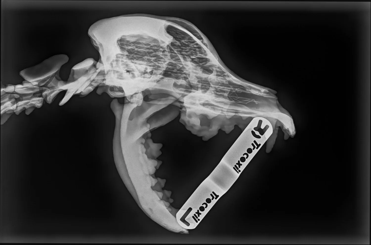
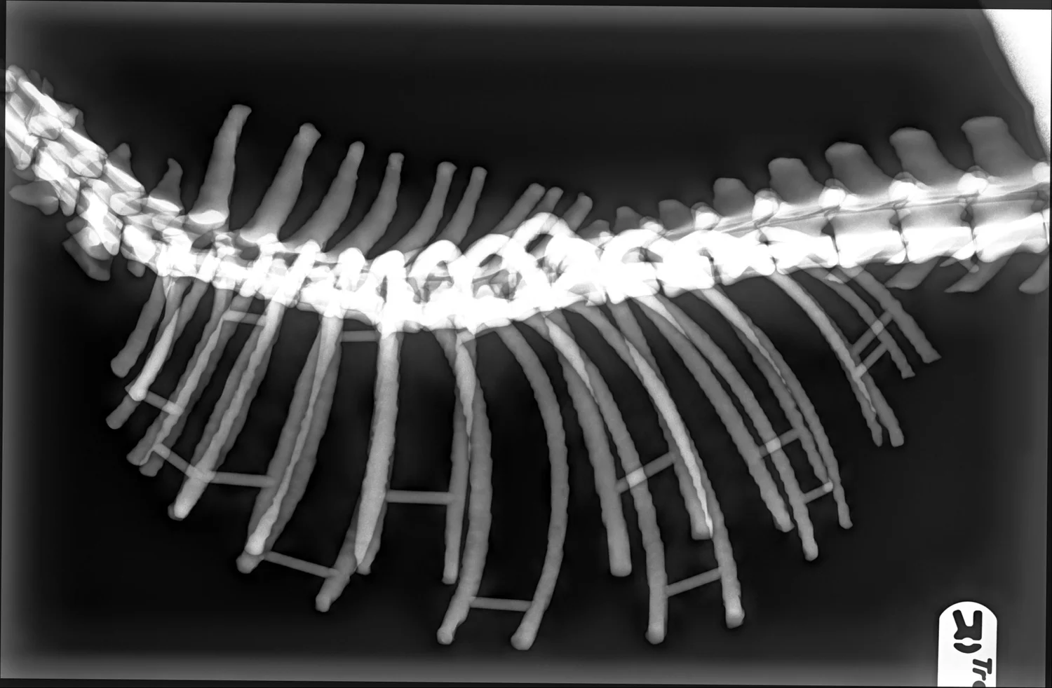
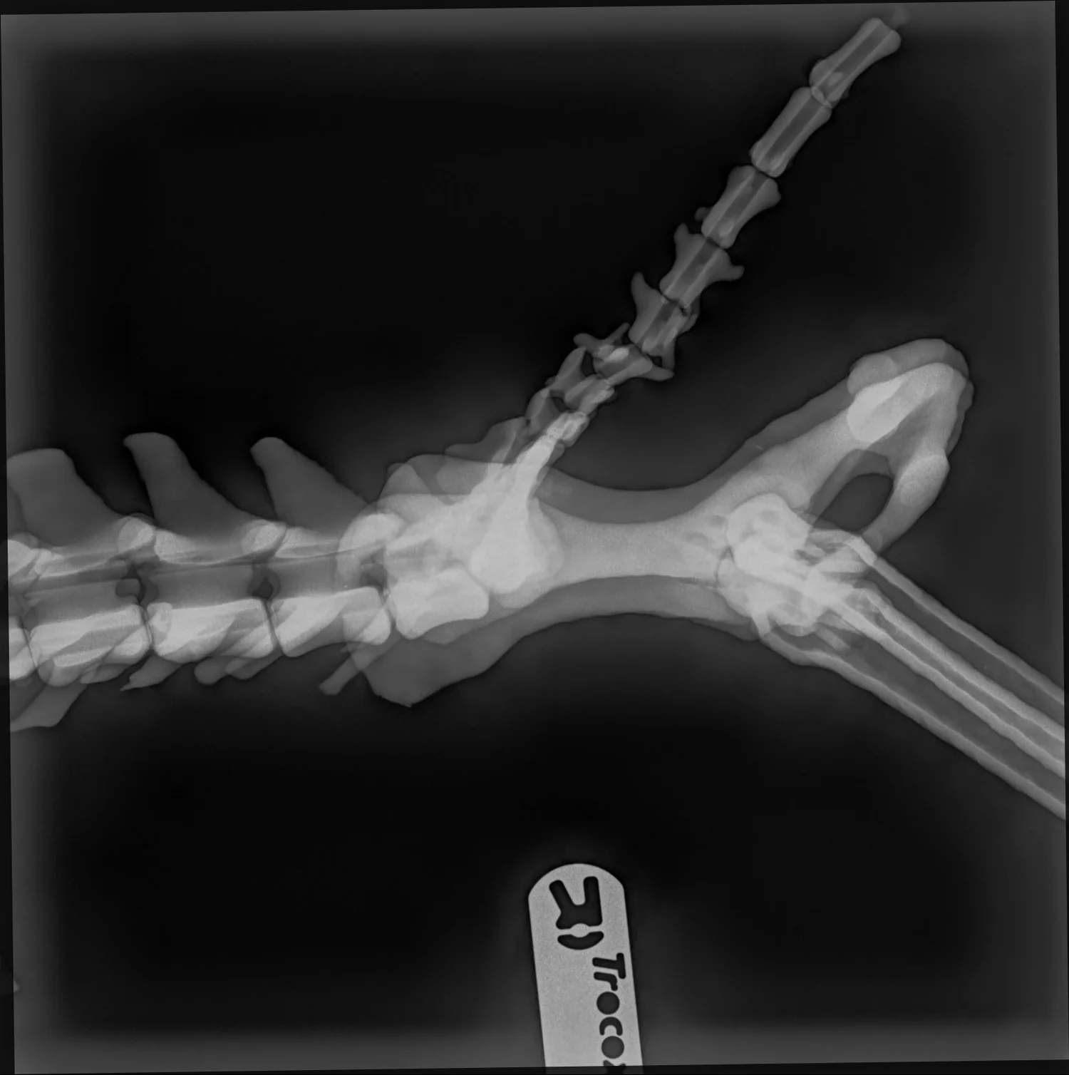
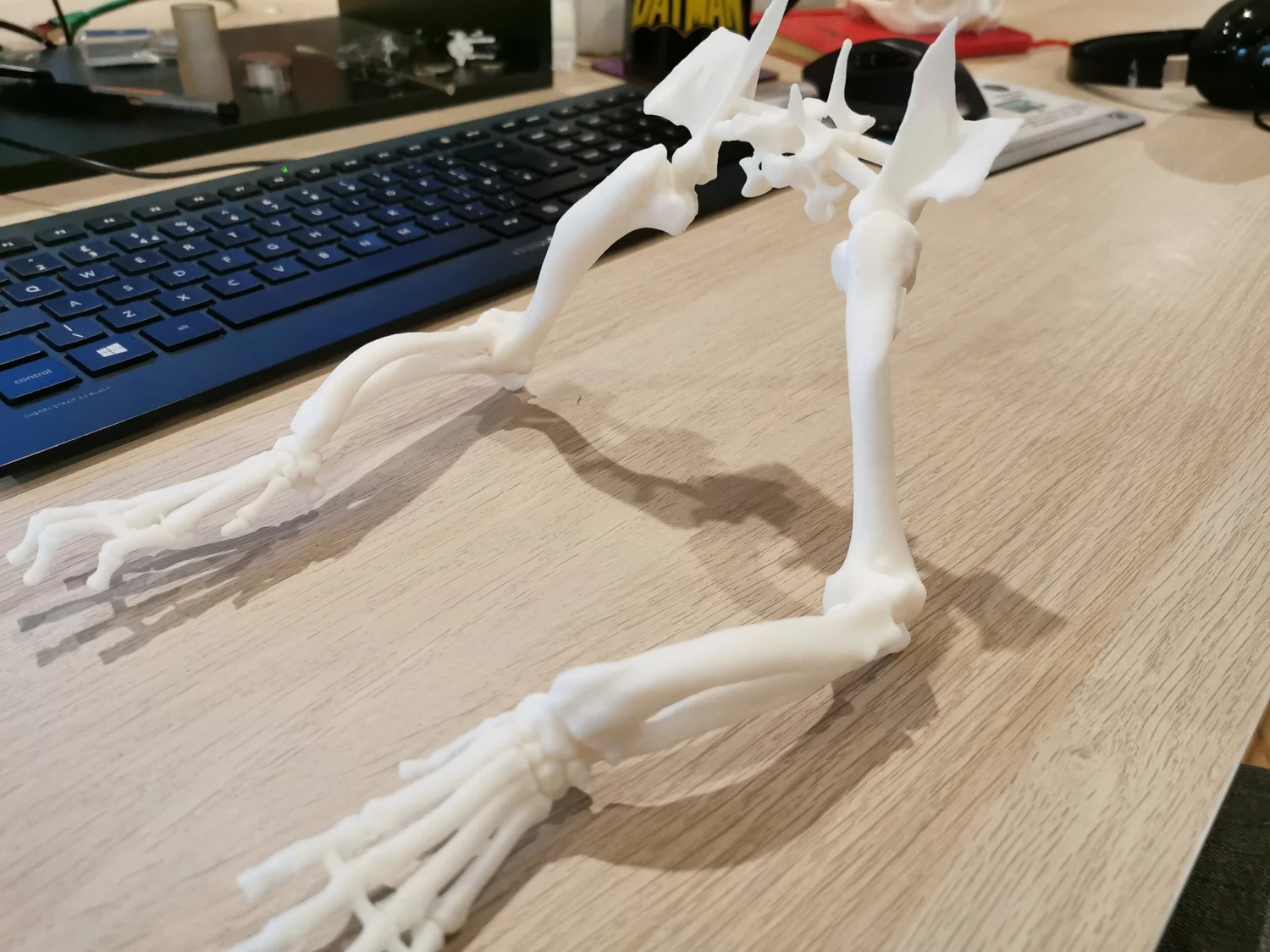
Canine simulator

X-ray opaque. Fully articulating. Created from real CT data. Do not buy it. Rent it.

Articulating canine anatomical model prepared for teaching and planning.
Radiograph of a canine skull and neck. Radiograph of a canine chest, rib cage and spine. Radiograph of a canine pelvis.

The model can be imaged and positioned so teams can rehearse a procedure before the patient is on the table.

Request a rental

Services

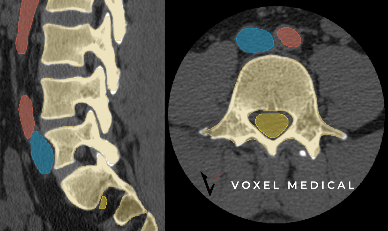
Segmented spine study with highlighted vertebral regions.

01

Anatomical modelling

CT and MRI data can be converted into high-precision 3D anatomical models for orthopaedics, trauma, cardiac work and oncology. A model can follow the patient scan closely, or it can be an idealised version that reduces artefacts and supports more general planning.

Order an anatomical model

02

Engineering

Articulations, fluid paths and other mechanical features can be added so a model or phantom behaves more like the living anatomy. Teams can plan or teach on a system that gives feedback, rather than on a passive static print.

Engineering consultation
Printed canine skeleton model on a workbench.
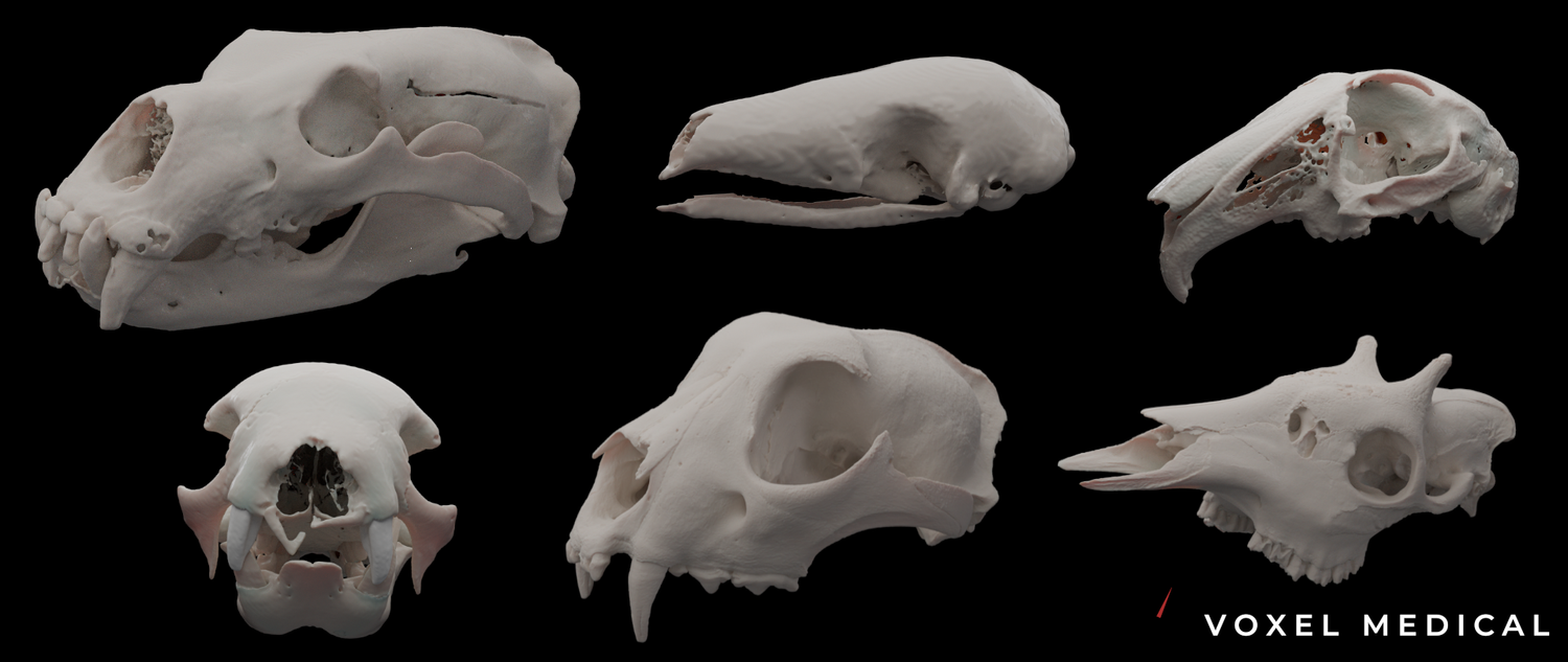
A row of mammal skull models used for comparative anatomy.

03

Educational models

Models for veterinary and zoological education show the biomechanics and structure of uncommon animals. Zoos, universities, museums and researchers use them for public teaching, comparative anatomy and specialist care of non-domestic species.

Order an education model

04

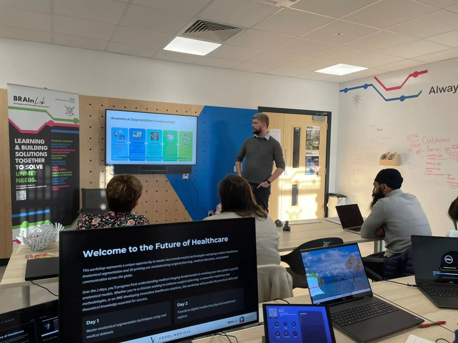
Segmentation and printing training

Training covers segmentation of anatomical scans, then printing and finishing of the parts. Courses run at basic, intermediate and advanced levels.

Enquire
A training session with a presentation and laptops on the tables.
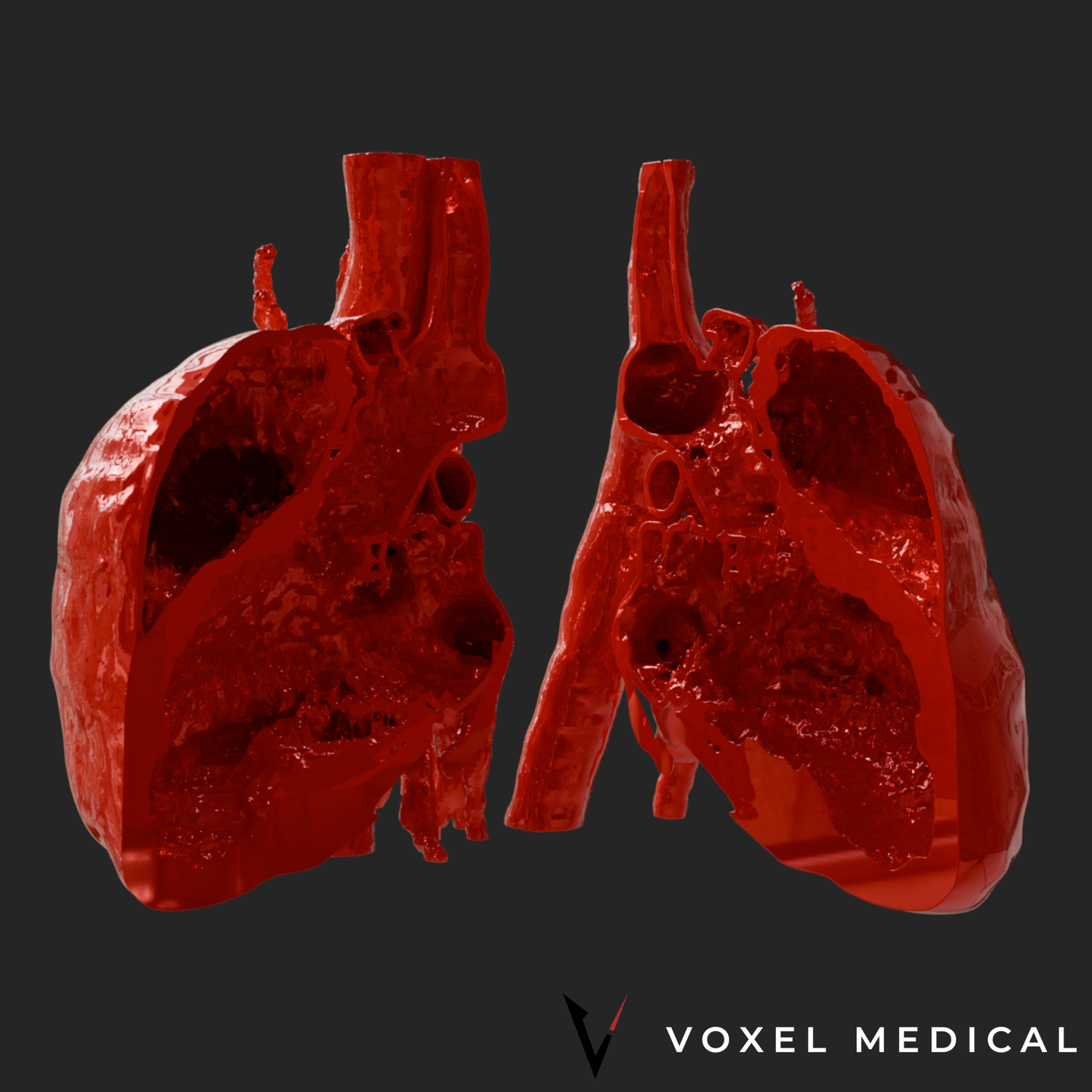
A canine heart model used to explain anatomy.

About

Models that help a team see the plan

Osseva builds anatomical models for surgeons, veterinary specialists and educators. Patient imaging can become a physical reference for planning, consent conversations and teaching. The same workflow covers domestic animals and less familiar species.

These models support planning and education. They are not medical devices and they do not replace clinical judgement.

Talk through an idea

Projects

Recent modelling work spans companion animals and zoological specimens.

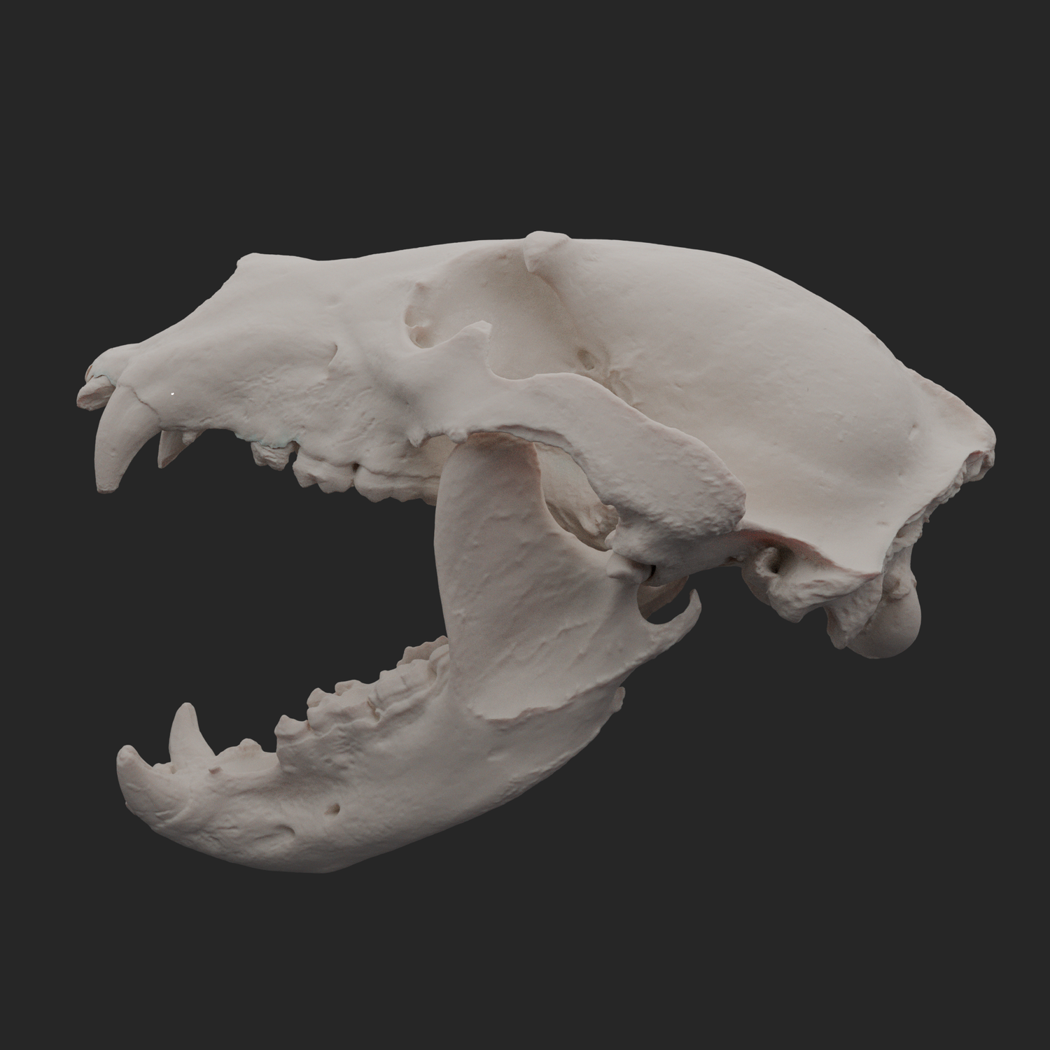
Visualisation of a skull model.

Brown bear skull

A reference model for comparative anatomy and zoological teaching.

Segmented canine spine.

Canine articulating spine

A movable spine for planning and demonstration.

Canine jaw radiograph.

Sloth dental

Dental anatomy prepared as a teaching reference.

Canine limb and body model.

Canine radius malformation

A patient-specific limb study for pre-operative planning.

Canine heart model.

Canine heart

A cardiac model for explanation and rehearsal.

Several skull models side by side.

Bobcat and canine skulls

Skull studies for lynx rufus and domestic canine anatomy.

FAQ

What is a patient-specific model?

It is a physical model made from that patient’s CT, MRI or CBCT DICOM data, so the shape follows the scan rather than a generic textbook specimen.

How do surgeons use one?

Teams use it to rehearse an approach, check implant fit and explain the plan. The aim is a shorter, clearer time in theatre.

Can it help with consent?

Yes. A model gives an owner or a student something to hold while the procedure is explained.

What files are needed?

DICOM from CT, MRI or CBCT. A clinic sends the imaging through a secure transfer, then the relevant anatomy is segmented.

How long does it take?

Turnaround is usually from about 24 hours to one week, depending on the anatomy and the finish.

Which materials are used?

Rigid plastics, flexible materials, silicones and gels. Some options are biocompatible. The choice follows how the model will be handled or imaged.

Which regions can be modelled?

Orthopaedics, cardiology, maxillofacial work and oncology are typical. Veterinary models cover dogs, cats and exotic species.

Is the model a medical device?

No. It is for planning and education. It does not diagnose, treat or replace the clinical decision.

How does a clinic start?

Use the form on this page. Describe the case, the imaging you have, and whether you need a static model, an articulating model or a rental simulator.

Schedule a conversation

Speak with an anatomical modelling specialist and talk through the case. This form stays on the page.

Thank you. Your note stays on this page and is not sent anywhere.